Presented by: Tom L. Rosamond, MD, MSc, FAHA, FACC, FASE
Dr. Thomas Rosamond is Board Certified in Cardiovascular Disease. Dr. Rosamond is the Division Chief of Cardiovascular Imaging and Medical Director of Non-Invasive Imaging Services at The University of Kansas Health System. He earned his Postdoctoral Fellowship in Cardiovascular Magnetic Resonance Imaging at Washington University School of Medicine -Barnes Jewish Hospital in St. Louis, Missouri.
Beyond Traditional Imaging: A Paradigm Shift in Cardiac Risk Assessment
In the rapidly evolving landscape of cardiovascular diagnostics, technological innovations are fundamentally transforming our ability to detect and predict heart disease risk. Recent advancements in imaging techniques are challenging long-standing diagnostic paradigms, offering unprecedented insights into cardiac health. Healthcare professionals now have access to more precise, detailed, and actionable data, enabling better decision-making and ultimately improving patient outcomes.
The Limitations of Traditional Imaging
For decades, standard diagnostic techniques like D-Spec (single-photon emission computed tomography or SPECT) have been the cornerstone of cardiac risk assessment. However, these methods have significant limitations when it comes to providing a comprehensive and nuanced evaluation of heart health.
One of the major drawbacks of traditional imaging is the image resolution and sensitivity constraints, which can result in missed diagnoses, particularly in patients at high risk for cardiovascular events. For instance, subtle but critical cardiac abnormalities, such as early-stage ischemia or coronary artery disease, may not be adequately captured, leaving these high-risk patients undiagnosed and without appropriate intervention.
As a result, healthcare professionals face a challenge: while these traditional methods have their place, they may not be sufficient for identifying patients who are at the highest risk of severe cardiac events.
PET CT: A Diagnostic Game-Changer
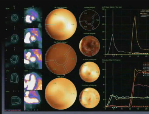
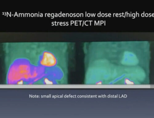
The advent of nitrogen ammonia positron emission tomography (PET) combined with computed tomography (CT) represents a revolutionary leap in cardiac imaging. This cutting-edge technology offers a dramatic shift in how we assess heart health, providing insights that were once out of reach with older imaging modalities.
Key Features of PET CT Imaging:
- Superior Signal-to-Noise Ratio: PET CT imaging dramatically enhances image clarity and diagnostic precision, making it possible to detect even subtle cardiac abnormalities that were previously undetectable with conventional imaging.
- Rapid Examination: A full PET CT scan can be completed in as little as 30 minutes, offering a time-efficient solution for patients and healthcare providers alike.
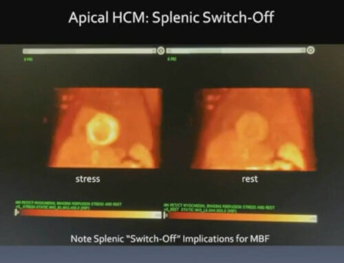
- Detailed Visualization: The technology’s ability to visualize coronary blood flow and myocardial tissue offers a level of detail previously unachievable, allowing for a deeper understanding of a patient’s cardiac health.
Key Advantages of PET CT in Cardiac Risk Assessment
1. Enhanced Risk Stratification
PET CT offers an advanced approach to stratifying cardiac risk, helping clinicians identify high-risk patients with greater accuracy. Through its ability to provide quantitative data on cardiac perfusion defects and its capacity to identify ischemic areas, PET CT allows healthcare providers to make more informed decisions regarding patient treatment. This leads to early intervention and the possibility of personalized treatment strategies, significantly improving outcomes.
2. Comprehensive Diagnostic Capabilities
PET CT provides a comprehensive diagnostic profile of a patient’s cardiovascular health, offering a detailed assessment of coronary blood flow. It allows clinicians to assess the reversibility of ischemic tissue and measure the extent and severity of cardiac defects. This level of detail supports precise targeting of medical or interventional treatments, optimizing the therapeutic approach for each patient.
Clinical Implications
A growing body of evidence and clinical case studies highlights the transformative potential of PET CT technology in cardiac risk assessment. In these studies, PET CT has demonstrated its ability to:
- Detect evolving cardiac risks within weeks, providing critical insights into the progression of coronary artery disease.
- Identify patients at imminent risk of serious cardiac events, enabling more proactive management of cardiovascular health.
- Guide the development of more targeted interventional strategies, from pharmaceutical treatments to surgical procedures, tailored to the individual needs of the patient.
As these case studies reveal, PET CT can be a game-changer for healthcare providers in managing heart disease and improving patient survival rates.
Navigating the Future of Cardiac Care
As healthcare professionals, it is essential to embrace these technological advancements. PET CT offers a more nuanced and precise approach to cardiac risk assessment, moving away from traditional binary diagnostic models. By integrating this advanced imaging technique into routine diagnostic protocols, healthcare providers can achieve a higher level of accuracy, leading to better patient care and more effective interventions.
Recommendations for Clinical Practice:
- Integrate advanced imaging techniques into diagnostic protocols: The integration of PET CT into routine clinical practice can lead to better outcomes for patients, especially those at high risk for heart disease.
- Stay informed about emerging cardiovascular imaging technologies: Technological advancements continue to emerge at a rapid pace. Healthcare professionals should stay updated on the latest tools and technologies to provide the most cutting-edge care to their patients.
- Consider the limitations of traditional diagnostic methods: While traditional imaging techniques still have their place, they should be supplemented with more advanced methods like PET CT to improve diagnostic accuracy and patient outcomes.
The leap forward in cardiac imaging provided by PET CT is not just about capturing clearer images; it’s about transforming our approach to diagnosing and treating heart disease. By offering more detailed, precise, and actionable data, PET CT enables healthcare providers to detect risks earlier, make better decisions, and implement personalized treatment strategies. As we move toward a more patient-centered and technology-driven model of care, embracing these innovations is crucial for improving patient outcomes and saving lives.


